
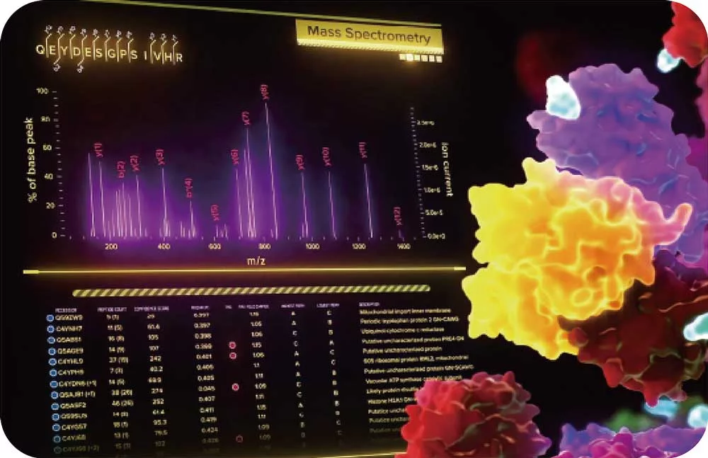
Traditional proteomic technology often relies on predefined panels or antibody-based enrichment,
which introduces bias—only known or expected proteins are detected.
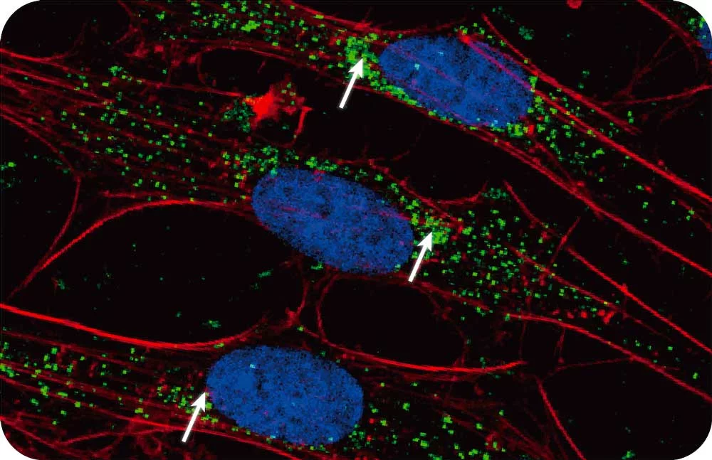
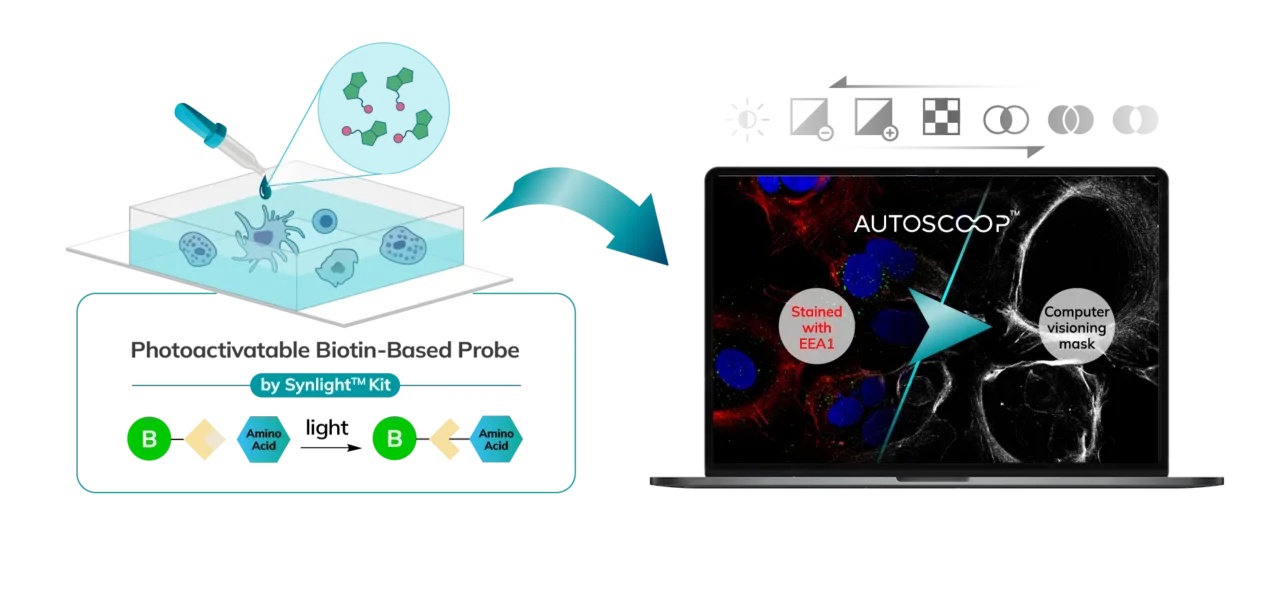
But Syncell’s Microscoop® labels and analyzes all proteins in a specific region of interest
without pre-selection, allowing mass spec to capture low-abundance or rare proteoforms, and reveal
otherwise unknown protein constituents with higher dynamic range and specificity than laser
microdissection or proximity labeling methods.
Mass spec alone doesn’t provide spatial information, but combining it with Syncell’s microscopy-guided photo-biotinylation technology enables researchers to link spatial location with the entire local proteome, allowing you to:
By restricting proteomic analysis to defined cellular or subcellular regions, our proprietary opto-proteomic platform improves:
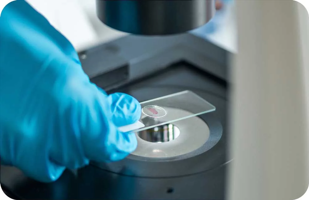
Microscoop® works with FFPE and fresh frozen tissue, preserving the integrity of archived clinical samples. This opens MS-based insights in areas where methods like proximity labeling cannot operate:
See Biology Like Never Before

Our advanced technology enables precise localization of proteins within cellular compartments, revealing intricate molecular interactions and pathways.

Explore the proteome without preconceived hypotheses, identifying novel biomarkers and therapeutic targets through comprehensive spatial analysis.

Integrate high-resolution imaging with proteomic data to correlate morphological features with molecular expression patterns.

Streamline your research with our end-to-end automated solutions, from sample preparation to data analysis and visualization.
This streamlined process is designed to deliver comprehensive and actionable data from complex biological samples, ensuring high quality and resolution at every stage.
 >
>
Precise tissue handling and preparation, followed by high-throughput data acquisition, ensures the integrity of the proteome for downstream analysis.
 >
>
Raw data is rigorously filtered, normalized, and aligned using proprietary algorithms to maximize feature detection and ensure accurate quantification.
 >
>
In-depth statistical analysis is performed to identify differentially expressed proteins and quantify their abundance across various sample cohorts.
 >
>
The resulting protein lists are mapped to functional pathways and processes, generating a clear, comprehensive report for actionable biological insights.